Oral Lichen Planus
a,b) Reticular, and c) plaque-like lesions of oral lichen planus in the same patient.
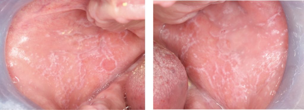
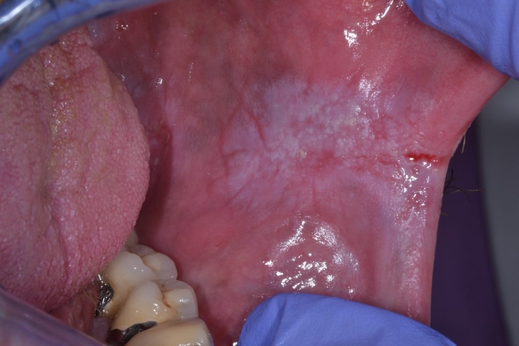
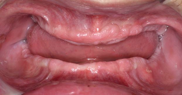
Bilateral atrophic/erosive oral lichen planus of the buccal mucosa
Bilateral atrophic/erosive oral lichen planus of the buccal mucosa
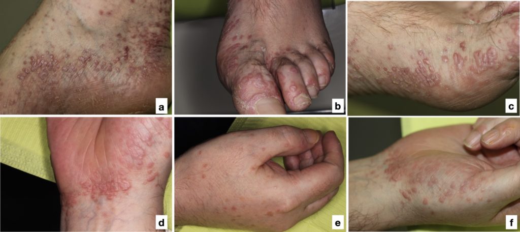
Cutaneous involvement in a patient with oral lichen planus (Figure 1). Papular lesions in a-c) feet and d-f) wrist and hands.
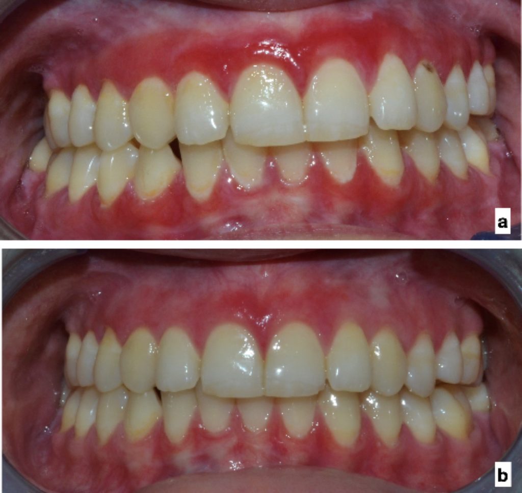
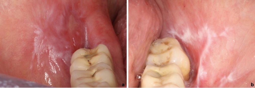
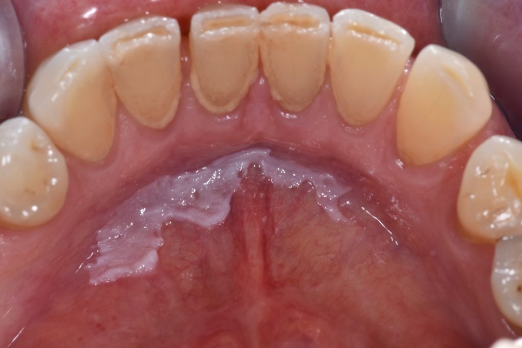
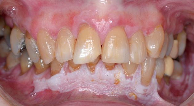
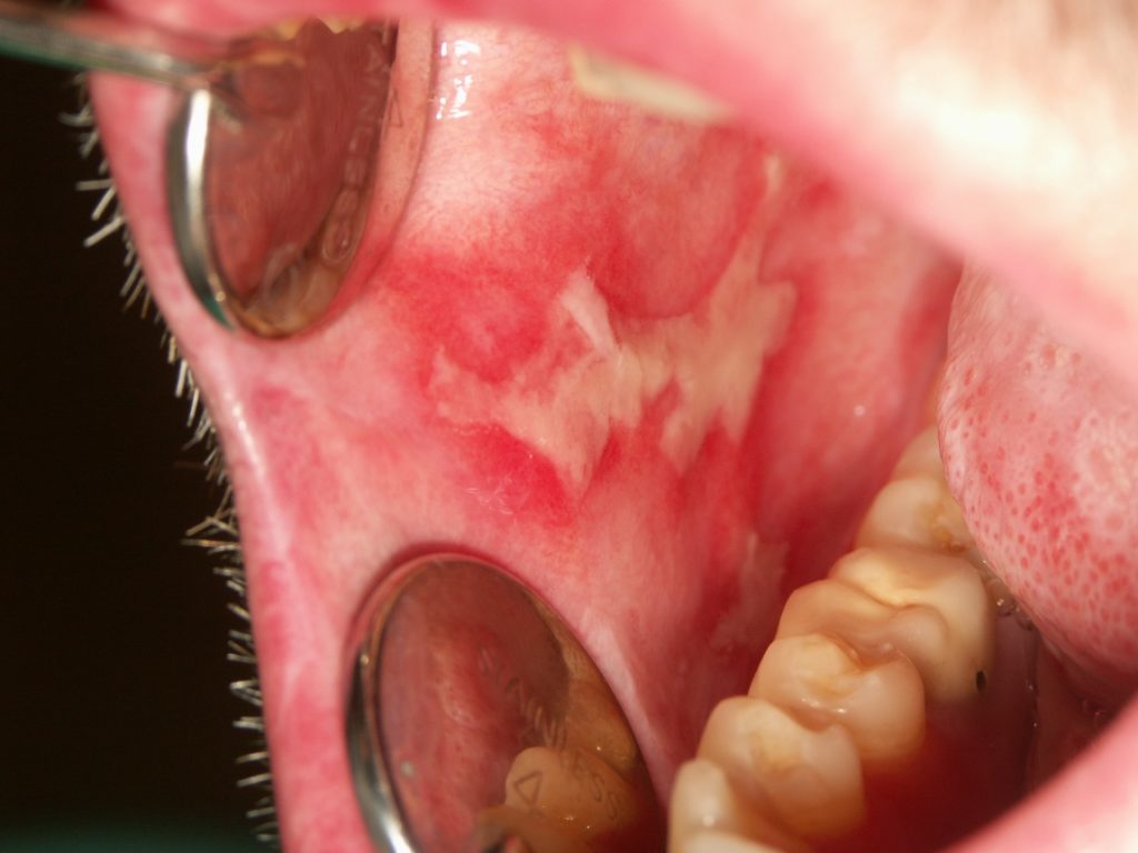
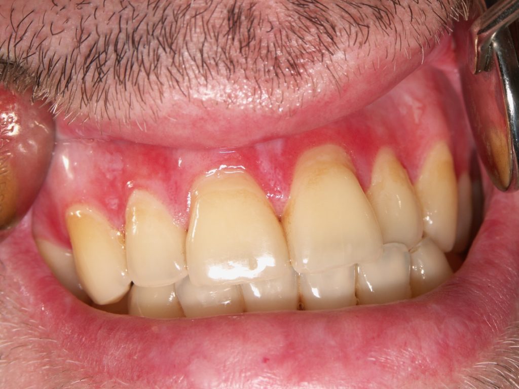
Desquamative gingivitis in a patient with oral lichen planus a) before and b) after treatment with 0.3% triamcinolone acetonide in custom trays.
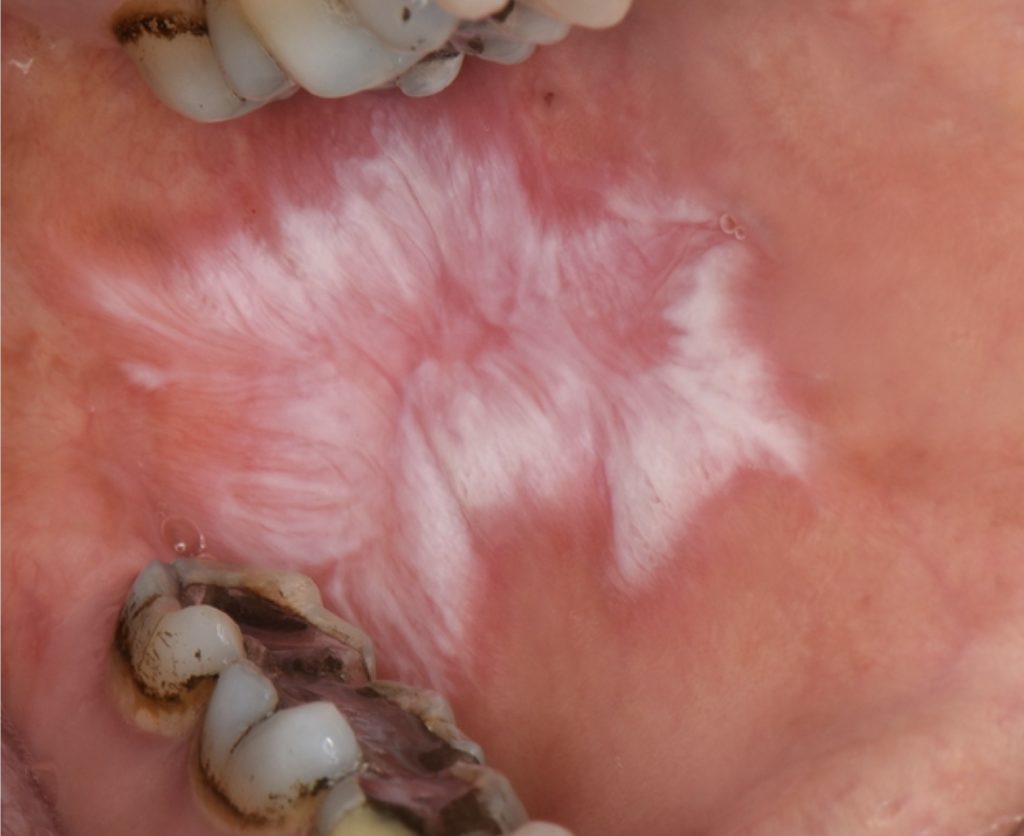
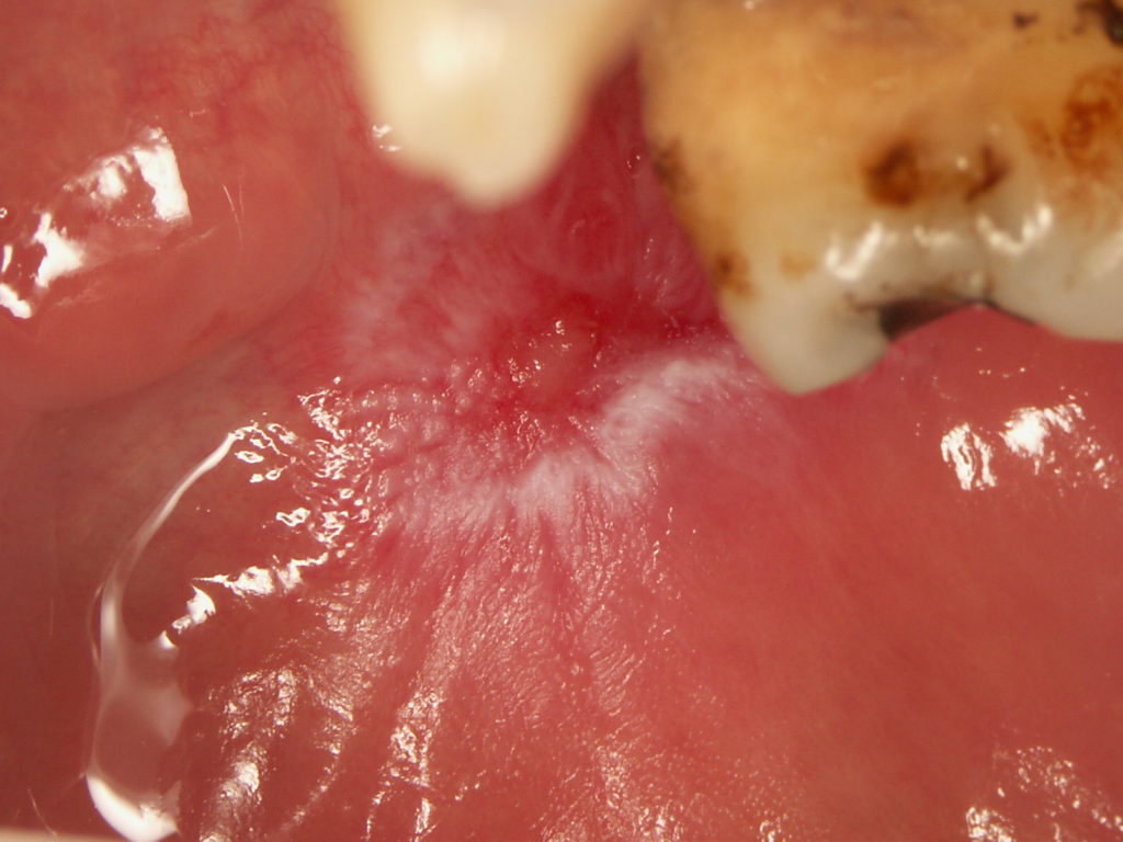
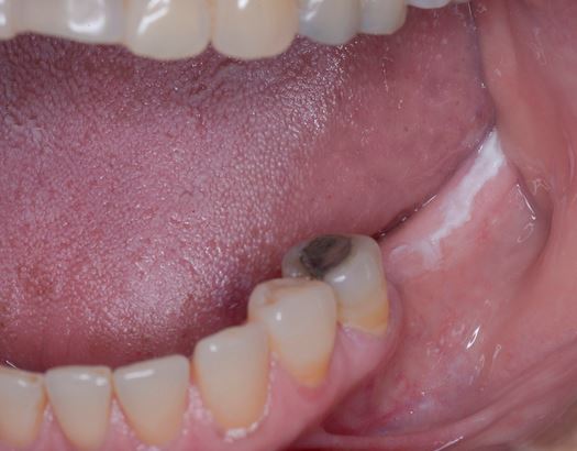
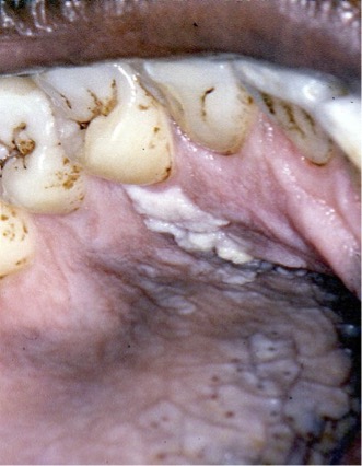
Oral lichenoid lesion in close contact with amalgam dental restoration.
Lip lesion in DLE presenting with central atrophy surrounded by radiating hyperkeratotic striae (courtesy of Professor Ivan Alajbeg)
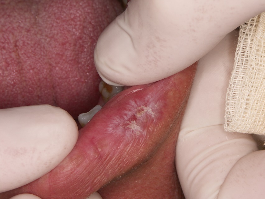
Oral mucosal lesion of DLE presenting with central atrophy and erosion surrounded by radiating hyperkeratotic striae (courtesy of Professor Ivan Alajbeg)
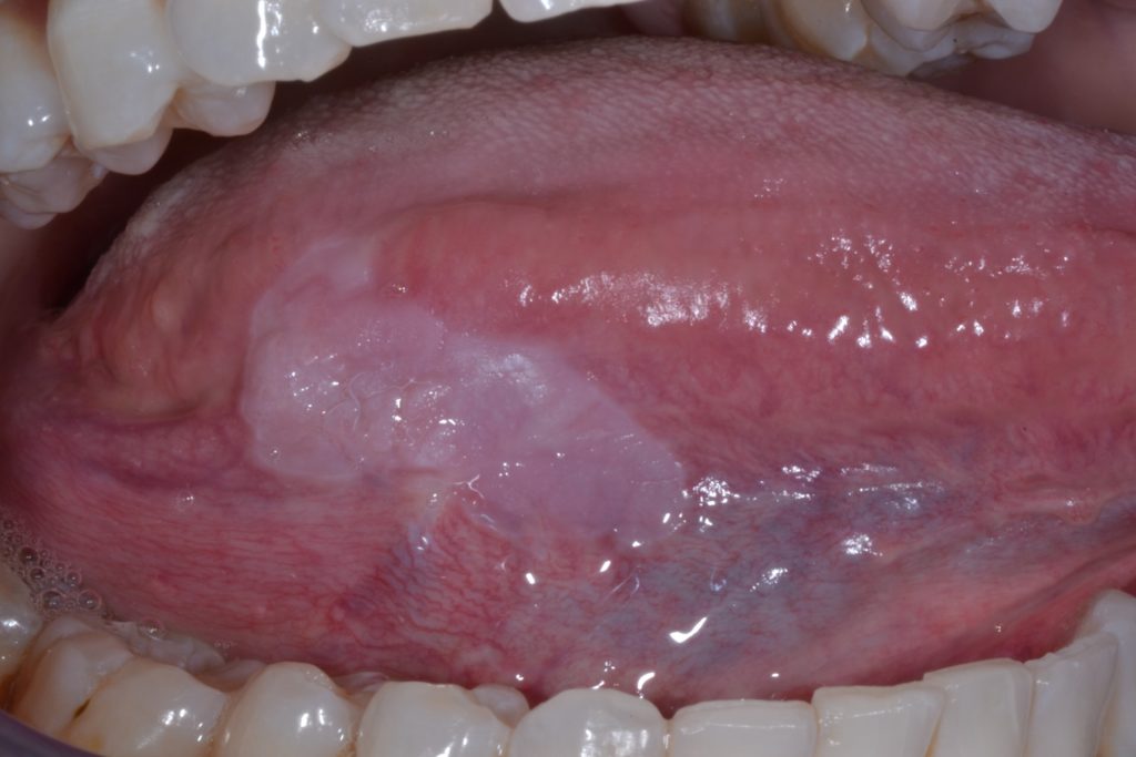
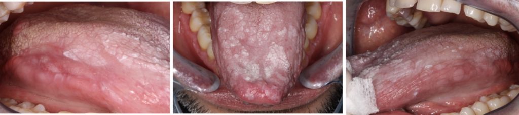
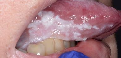
Homogenous Leukoplakia of the mid right lateral tongue
Non-homogenous leukoplakia involving the right lateral and ventral surfaces of the tongue
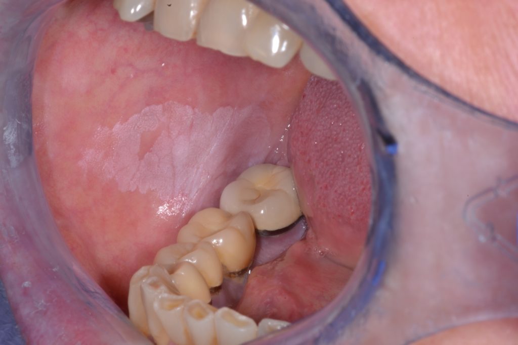
Homogenous Leukoplakia of the right buccal mucosa
Non-homogenous leukoplakia with verrucous and nodular components affecting the left buccal mucosa.
Non-homogenous leukoplakia involving the encompassing largely the right lateral tongue with more prominent keratotic areas seen, not exophytic.re 5: Non-homogenous leukoplakia involving the encompassing largely the right lateral tongue with more prominent keratotic areas seen, not exophytic.
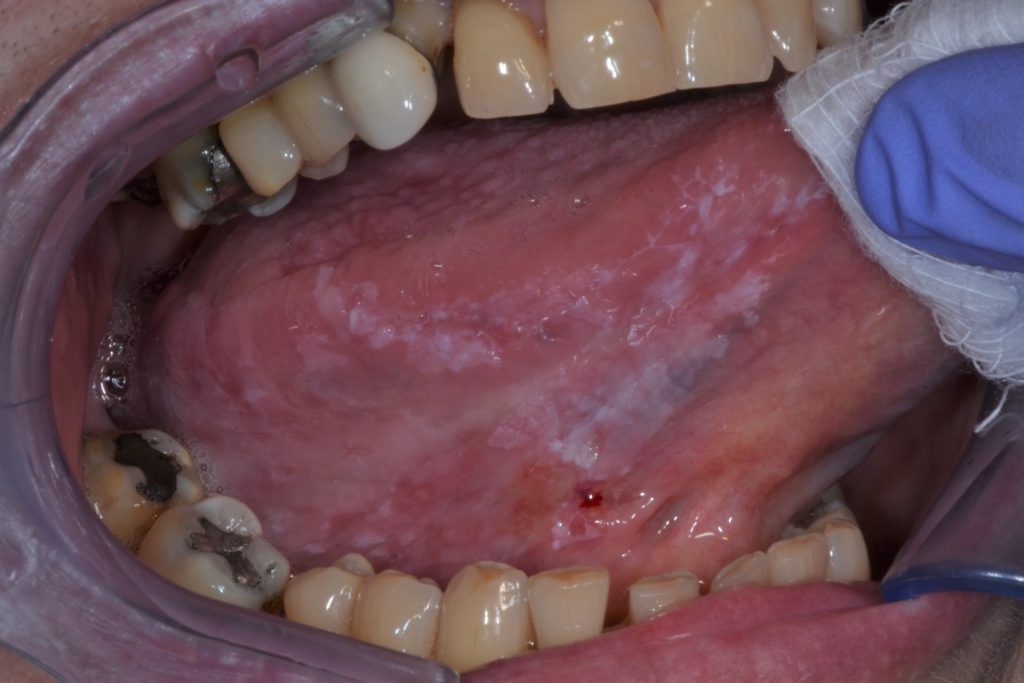
Largely Homogenous Leukoplakia of the floor of mouth
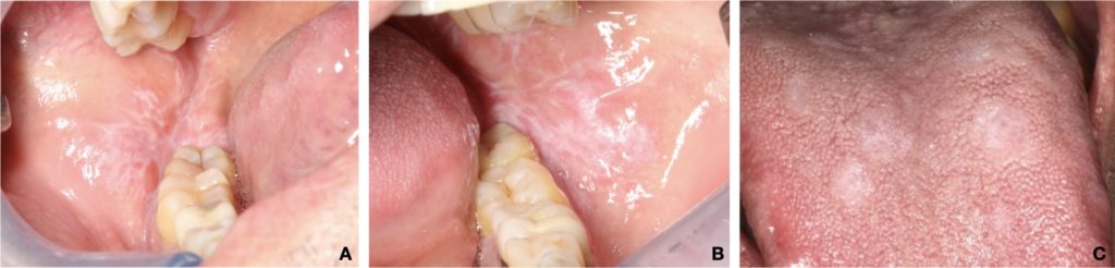
Mild PVL affecting the edentulous ridges
A dense multifocal area of PVL on the encompassing the right lateral tongue
A dense multifocal area of PVL on the encompassing the right lateral tongue
PVL affecting the lower left posterior edentulous ridge
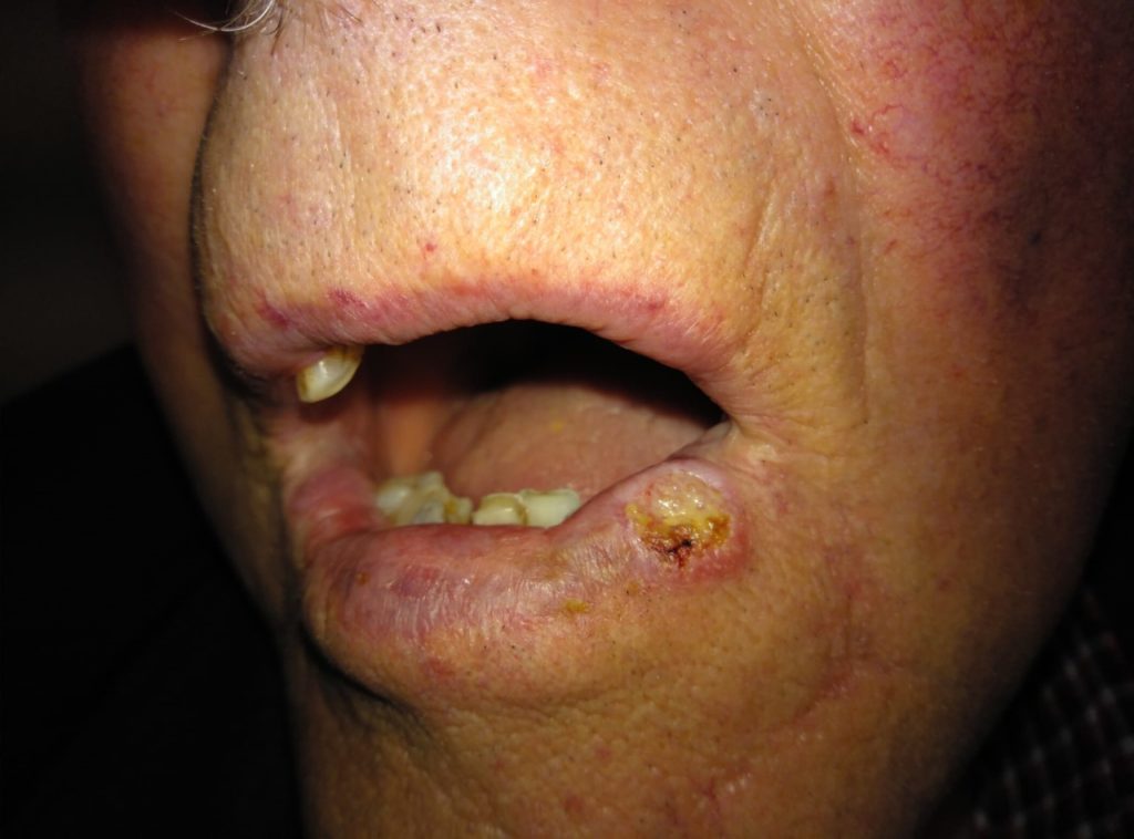
Squamous cell carcinoma with background actinic cheilitis involving the lower lip. The lips appear dry and cracked with loss of the vermilion border. An ulcer with everted edges and indurated on palpation is noted on the left lower lip.
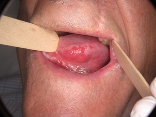
Figure A
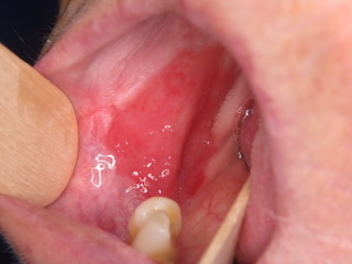
Figure B - Widespread Erythroplakia homogenously affecting the left lateral border of the tongue and homogenous erythroplakia homogenously affecting the right buccal mucosa posteriorly.
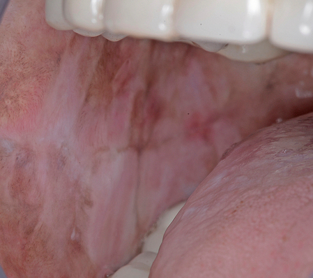
Figure A

Figure B
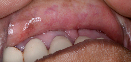
Figure C
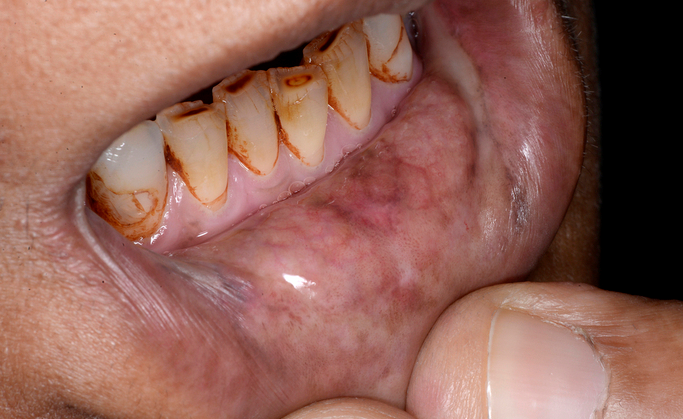
Figure D - Figures A-D: Showing Blanching of the left and right buccal mucosa with leukoplakia and some evidence of fibrous banding visible, more prominent on the right buccal mucosa. Loss of pallor is evident on the upper and lower labial mucosa.
Ulcerative lesion involving the buccal mucosa in a patient with cGVHD
Gingival involvement presenting with erythema and striations in a cGVHD patient
Palatal Lesions in Reverse Smokers
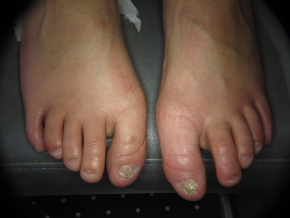
Dystrophic nails in Dyskeratosis congenita affecting the toes nails.
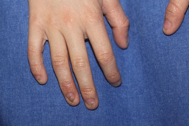
Dystrophic nails in Dyskeratosis congenita affecting the finger nails.
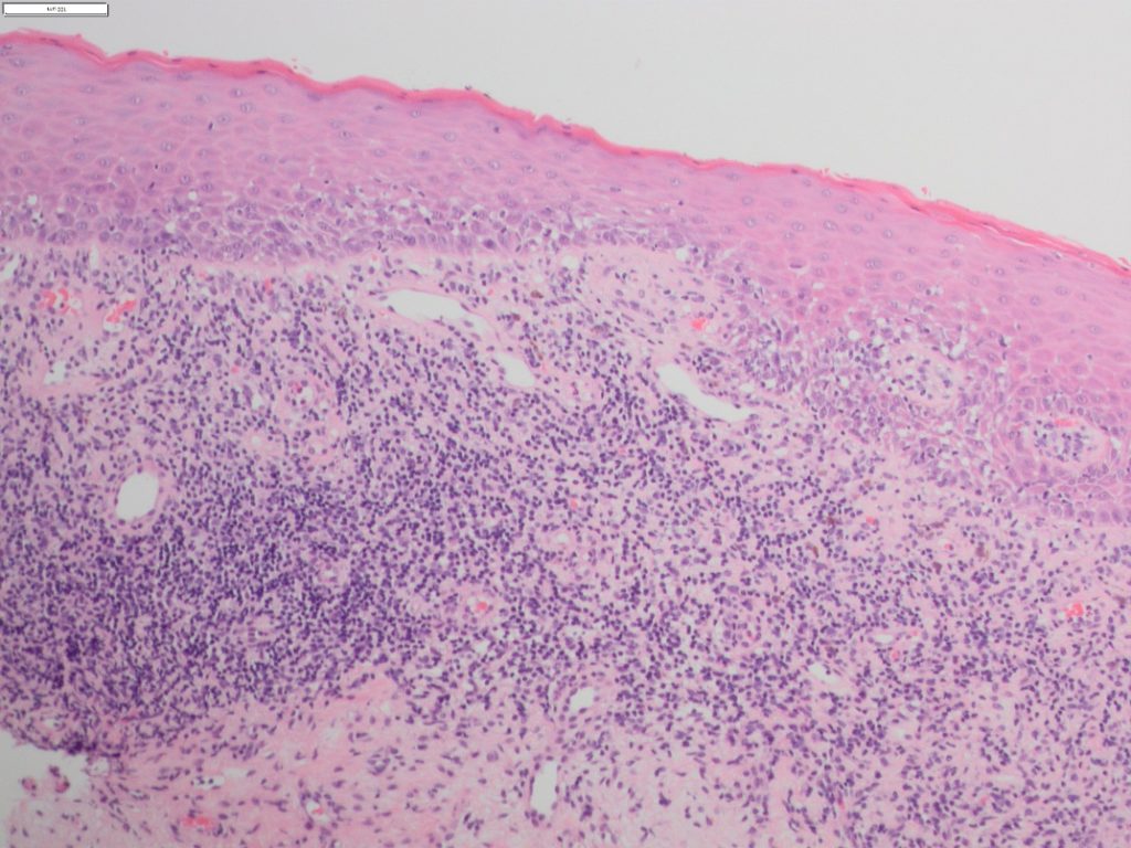
A photomicrograph showing the typical histological characteristics shared amongst the oral lichenoid lesions including hyperkeratosis, epithelial atrophy with basal cell degeneration and a dense band-like lymphocytic infiltrate within the lamina propria.
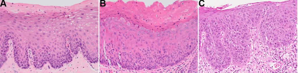
Examples of A) mild dysplasia demonstrating hyperchromasia and focally increased nuclear to cytoplasm ratio limited to the basal and parabasal layers, B) moderate dysplasia highlighted by disordered stratification and maturation affecting half the epithelial thickness, and C) severe dysplasia featuring full-thickness architectural and cytological atypia.